When you have completed review of this screen, please click the "Next
page" blue arrow for the second portion of this section.
When you have completed review of this screen, please click the "Next
page" blue arrow for the second portion of this section.
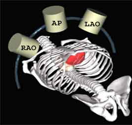
The AP view is not commonly
obtained because the spine sits in the middle of the cineangiographic
picture in this particular view and the coronaries lie directly in front
of it. The similarity in radiographic density between the spine and
the contrast-filled coronaries interferes with the quality of the images.
However, it is occasionally obtained when the location and severity
of a stenosis cannot be clearly defined in the conventional views.
The AP view may be of particular value in evaluating the ostium
of the left main coronary artery and the shaft of the mid LAD.
The AP view is of historical interest and goes back to the early
days of cardiac catheterization, The x-ray tube, image intensifier
and camera remained stationary, while the patient lay in a cradle
that was rotated from the RAO to the LAO projections. Cranial or caudal
angulation were not possible at those times. The AP view was often
the first "scouting" view.
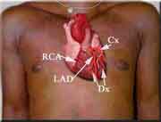
The central or "neutral" position of the camera is known as
the AP (antero-posterior) view, at which time the camera "looks" straight
down the patient's chest and heart. In other words, the X-ray camera
is directly above the patient's chest with the beam coming straight
up from the x-ray tube and perpendicular to the patient (below, left).
A camera's view of the patient's heart in the AP view is shown on
the right (below). The size of the heart has been purposely exaggerated
for purposes of illustration.

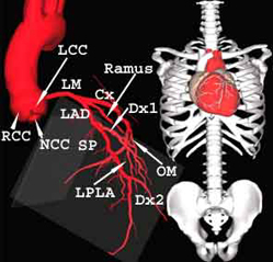
The aortic valve has three cusps. The RCC or right coronary
cusp, where the right coronary artery originates and the LCC or left
coronary cusp where the the left main (LM) coronary artery arises.
The NCC or non coronary cusp sits posterior to the other two cusps
of the aortic valve.
The LM divides or bifurcates into the left anterior descending
(LAD) and the Circumflex (Cx) coronary arteries. In many cases there
is a third artery that originates from the LM and travels between
the LAD and CX. This artery is known as the Ramus Intermediate (Ramus)
or Optional diagonal coronary artery. In the AP view the LAD runs
down the front of the heart along the side of the spine. The ramus
and diagonal (Dx) moves diagonally and away from the LAD and the spine
in this view. When more than one Dx is present, the first one is called
the first Dx or Dx1, the second is called Dx2, etc.
The septal perforators (SP) are smaller branches that come
off the LAD. They supply blood to the inter-ventricular septum.
The Circumflex (Cx), in the AP view, moves away from
the spine at nearly a 90 degree angle and then wraps around the left
atrio-ventricular groove to the back of the heart as was previously
described. The Cx gives off one or more obtuse marginal (OM) and left
postero-lateral branches (LPLA) that run downwards and away from the
AV groove. When more than one OM branches are present, the first OM
is called OM1, the second is called OM2, etc.
|